
3D NLS-graphy With Hunter 4025 Scanner May Be Considered Method Of Primary Diagnostics Of Rectum Cancer
Nowadays oncourology is the sphere where methods of three-dimensional NLS-graphy may also be widely applied. However until this day application of three-dimensional NLS-research of patients operated on urinary bladder tumor consisted in dynamic monitoring of organŌĆÖs condition in order to detect recurrent tumor and metastases at early stage. Introduction of threedimensional NLS-methods into clinical practice allow complete changing of point of view to this problem. We believe that this issue is really topical, because majority of surgically operated patients were subjected to traumatic transurethral resections.
Three-dimensional NLS-research with application of spectral-entropy analysis, carried out during surgical oncotomy, allowed us to detect additional tumoral neoplasms, not registered by two-dimensional NLS-research in 37% of patients. Application of threedimensional methods makes possible to specify extent of tumor process local spreading, control depth of urinary bladder wall resection and decrease risk of complication development during oncotomy.
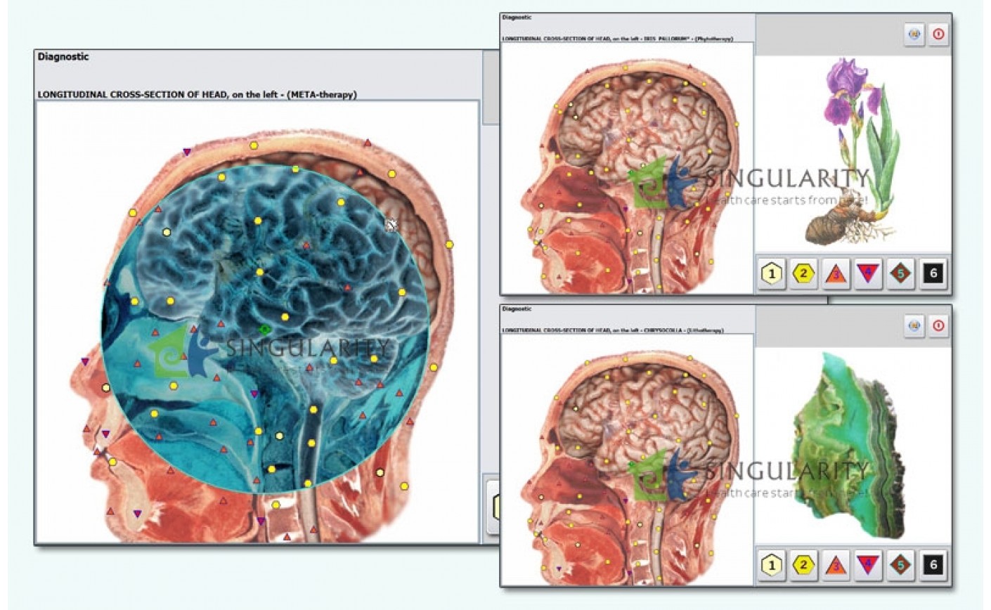
Diagnostics and morphological verification of rectum cancer does not present difficulties, as a rule. However evaluation of organŌĆÖs wall invasion degree is not always possible by standard methods of diagnostics. Traditional two-dimensional NLS-research is already widely used as diagnostics method of rectum recurrent cancer after organ extirpation. Nevertheless primary diagnostics of the disease by two-dimensional NLS-graphy is hindered due to several reasons. In the first place it is explained by the fact that at two-dimensional NLS-scanning rectum is visualized only partially (80% of whole organ surface area).
Application of three-dimensional NLS-graphy makes possible to differentiate accurately all layers of rectum walls, and thus to diagnose depth of tumor infiltration and identify stage of the disease, using spectralentropy analysis. This method helps to detect changed lymph nodes sized above 1.5 mm at metastatic disease of pararectal lymph nodes. During monitoring of carried out pre-operational radiotherapy three-dimensional NLS-graphy helps to detect accurately decreasing of tumor sizes, identify changes in its structure, related to medical pathomorphism, identify decreasing of pararectal tissues tumoral infiltration. Therefore three-dimensional NLS-graphy with hunter 4025 scanner may be considered method of primary diagnostics of rectum cancer. It allows therapist to solve the most important diagnostic issues, related to identifying of tumoral process length, extent of tumor local spreading and monitoring of carried out pre-operative treatment efficiency. At organ preserving operations three-dimensional NLS-graphy may be used as efficient method of recurrent tumors early diagnostics in anastomosis area.
What Is The Basis Of Trigger Sensors Effect - Metatron 4025 Hunter
For the first time distant effect of interaction between objects of animate and inanimate nature, in other words transfer of information impulses from a human to a device, was registered in experiments carried out by Valery Kravkov in the 20ŌĆÖ of the last century. Reactions of various semiconductor structures to human influence were studied under guidance of Professor Vyacheslav Togatov. It was proven by experiments that human brain can influence a sensor of a device without any wires.
What Is The Bioresonance? ŌĆō Metatron 4025 Hunter
Our world today has more illnesses than any other time in history. While typical medical science has come a long way in helping those who deal with long term illnesses, we are still lacking in just how we can prevent illness. The answer to filling in the gap between optimal health and the medical community just may be BioResonance.