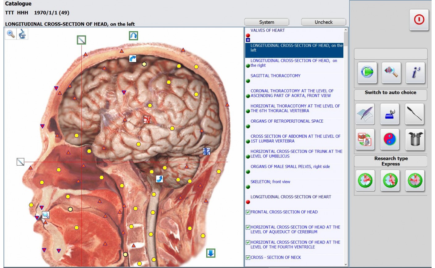
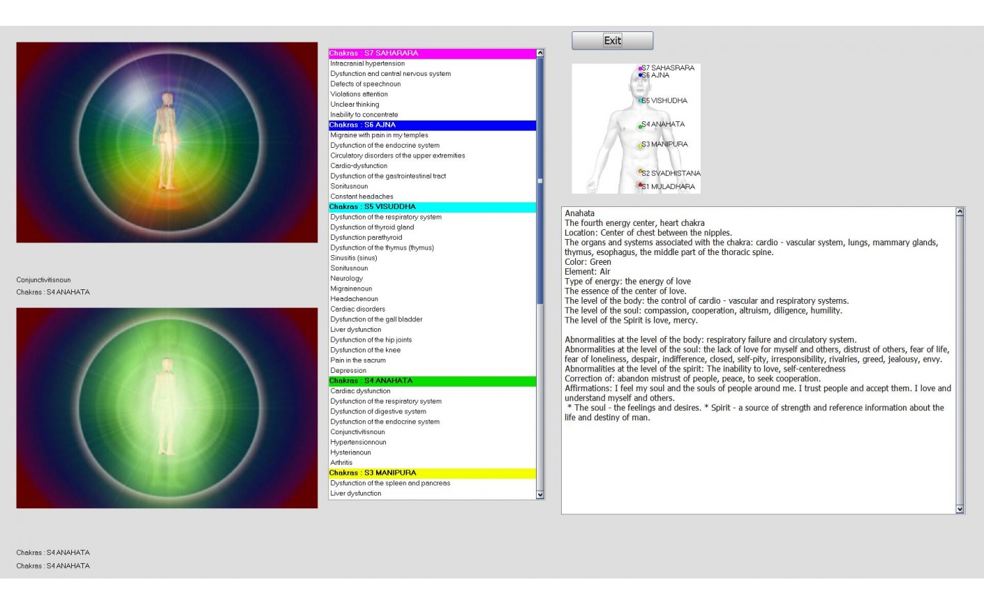
NLS-examination With Metatron 4025 Hunter For Liver Hemangioma
The most frequent unexpected finding after examination and the object of further diagnosis verification is liver hemangioma.
Hemangioma is a benign vascular neoplasm of liver (prevalence in population is up to 15%). Having heterogeneous internal structure, their visual picture may resemble cancer (especially at ultrasound and CT), which requires additional diagnostic investigations. In majority of cases hemangiomas are clinically asymptomatic.
Diagnostic criteria of hemangioma (according to MRI, CT and NLS data) are considered to be the following: it is never encapsulated, edematous, is drawn towards hepatic veins, sometimes its form is close to form of hepatic lobes. Its outlines may be of irregular form, but distinct. Dynamic study detects very slow growth.
At NLS-examination of metatron 4025 hunter hemangioma often visualized as hyperchromic (4 ŌĆō 5 points according to FleindlerŌĆÖs scale) neoplasm. However if there is corresponding fatty infiltration of liver present, hemangioma acquires hypochromeity and sometimes it is hard to differ it from cysts or metastases. Cavernous hemangioma is represented by hypo- and achromogenic areas (1 ŌĆō 3 points), which complicates its interpretation. Hyaline fissure, one of the most typical symptoms of hemangioma, not always can be revealed.
It should be noted that if there is possible hemangioma, puncture cannot be administered due to high risk of haemorrhage development. It requires dynamic monitoring of a patient and repeated examinations.


